
In addition to the clinical evaluation of the dental patient, imaging is a crucial diagnostic tool. A complete and comprehensive picture of the jaws and dental structures was made possible by the invention of panoramic radiography in the 1960s. Its wide-scale use throughout the 1970s and 1980s indicated a significant advancement in dental radiology. However, the inherent limitations of all planar 2D projections are magnifying, distorting, superimposing, and misrepresenting structures. This applies to intraoral and extraoral treatments, whether used separately or in tandem. The development of three-dimensional (3D) radiographic imaging has received much attention. Still, despite its availability, CT’s usage in dentistry has been constrained by issues with cost, accessibility, and exposure. Cone-beam computed tomography (CBCT), developed especially for imaging the dental-maxillofacial area (Christos Angelopoulos et al., 2012), marks a fundamental, radical transformation from a 2D to a 3D method of collecting data and image reconstruction. How does a cone beam ct work in a dental office for your better understanding?
Before going into a detailed discussion about how CBCT works, it would be better to have a bird’s eye view of this technology so that you can understand its working procedure easily.
Dental cone beam CT is a specialized form of x-ray apparatus used when standard dental or facial x-rays are insufficient. This technology lets dentists capture 3-D images of the tooth, soft tissues, nerve pathways, and bone in a single run. Since this scanner exposes patients to more radiation than standard dental x-rays, it is not frequently utilized. Cone beam computed tomography (CT) produces many images, commonly known as views, by moving an x-ray beam around the patient like a cone.Cone beam computed tomography is used to assess abnormalities of the jaws, teeth, bony features of the face, nasal cavity, and sinus. CBCT scans are relatively straightforward, more affordable, and require less complex software and hardware than CT scanners, yet they nonetheless generate equivalent or better pictures of hard tissue components of the skull and jaw.
Patients can be scanned using cone-beam technology in one of three positions: sitting, standing, or supine. Patients having physical limitations might not be able to use technology that necessitates them to lie supine since it takes up more space and has a bigger physical footprint. It is possible that standing units can’t be placed to a level where patients who use wheelchairs can use them. Seating units provide the most convenience. Nevertheless, people who are physically impaired or confined to wheelchairs may not be able to be scanned on fixed seats. Regardless of how the patient is positioned within the apparatus, the basic ideas behind image generation never change.
The four steps involved in creating a CBCT image are:
Depending on the below-mentioned benefits, cone-beam CT has replaced conventional spiral and helical CT as the most sophisticated radiographic methodology (Jabero, Marvin, Sarment, David, 2006):
However, even with the usage of this technology, inaccuracies of 1.5 mm and 1 mm in horizontal and vertical dimensions have been observed, indicating that the accuracy of the CBCT approach still needs to be discovered.
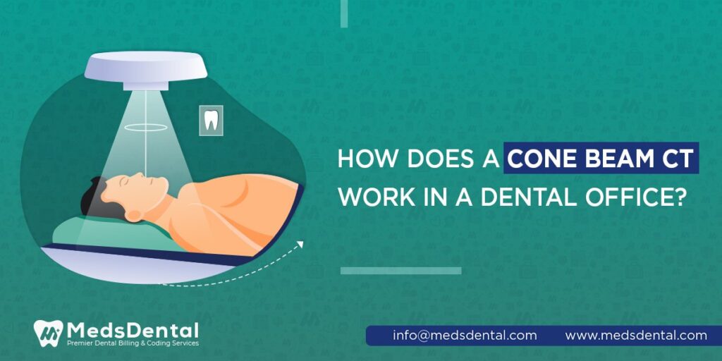
Based on the CBCT scanner type, the person may be requested to lie down on the examination table or sit in the examination chair. The targeted area should be centered in the beam when the dentist adjusts the patient. The patient will be instructed to maintain extreme stillness because the x-ray source and detector rotate around their head and face for a maximum 360-degree spin.
This usually takes between 20 and 40 seconds for a total volume, also known as a full mouth x-ray, in which the whole mouth and dental components are photographed. A regional scan, which concentrates on a particular location of the maxilla or mandible, takes less than 10 seconds.
It is a noteworthy point that before starting the procedure;
A kind of radiation known as X-rays exists on the electromagnetic spectrum. It is analogous to an invisible light that can pass through the bone and soft tissue to produce a picture that reveals underlying patterns. The intense X-rays show what is below the layer in grayscale with variable degrees of absorption. In a cone beam CT scan, the C-arm or gantry revolves 360 degrees around the patient’s head while taking several images from various angles combined to produce a single 3-D image. The rotating C-arm or gantry has an x-ray producer fixed on one end and a detector attached to the other. The detector can produce 150 to 200 high-resolution two-dimensional photographs in a single cycle. These images are then electronically combined to create a 3-D image, which can give your dental surgeon essential details about the condition of your teeth and craniofacial structure.
According to the area of focus, picture quality, patient size, site of the region of interest, and manufacturer settings, the radiation exposure from a cone-beam CT of the jaws can range from around 18–200 µSv.
Dentists frequently employ a Dental Cone Beam CT to design orthodontic and dental implant treatments. Cone Beam CT imaging is indeed helpful in more complicated situations that include:
CBCT has some drawbacks, including:
Due to their demanding schedules and daily patient load, dentists need help managing the billing process effectively. Thus, MedsDental Dental Billing Company provides dependable billing services to help dentists feel less stressed. Due to our extensive expertise in billing, we can streamline your income while lowering AR days and boosting cash flow. Our knowledgeable staff applies legitimate codes to the CBCT services you provide to the patients. We submit the accurate claim to the insurance company for payment after preparing it with the proper coding of the cone beam computed tomography service. We use the most recent software while abiding by HIPAA regulations, which allows the entire billing process to function quickly and safely. For your practice and billing procedures, our company is a full-service solution.
© MedsDental. All rights reserved 2026. Powered by MeshSq.